Retina / Retina
What is the retina?
The retina is a sensory organ in the eye, where images we see are projected and processed in the form of a nerve impulse that travels along the optic nerve.
It works like the film in a camera. It is the innermost layer at the back of the eye. The images enter through our pupil, pass through the crystalline lens and are focused by the retina. It then converts them into electric signals and sends them to the brain via the optic nerve.
The most common retina disorders are macular degeneration (AMD), diabetic retinopathy and venous thrombosis. Retinal detachment, epiretinal membrane and macular holes are also fairly common, and are all treated by surgery.
As for the vitreous body, this is a gelatinous substance that fills the space between the crystalline lens and the retina, maintaining the eye’s shape. This vitreous gel may deteriorate and cause condensation or clumps that can project shadows onto the retina and produce the appearance of infamous “floaters”, or myodesopsia.
There are a number of disorders that affect the vitreous body, such as vitreous detachment and loss of transparency, which can be caused by vitreous haemorrhage.
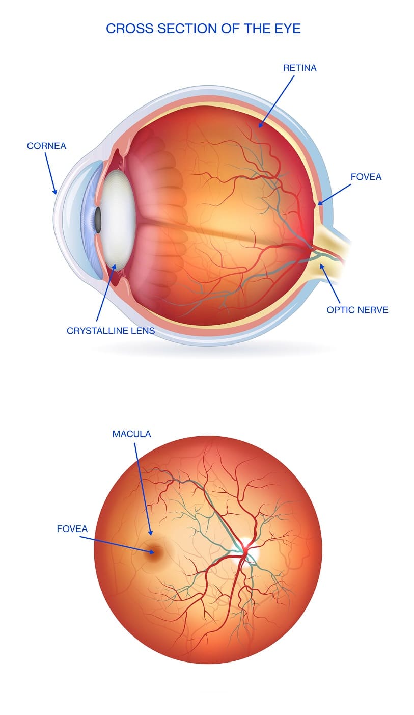 Parts of the eyeball
Structure of the retina
The retina is made up of a network of nerve cells that travel from the brain to the optic nerve. The most important thing is to first define the fundamental parts of the eye and understand how they work, as many retinal disorders are the primary causes of blindness in Spain, which is why it is essential to detect them early.
We will now outline the anatomical distribution of the fundus of the eye:
→ Optic disc
→ Retinal artery and veins
→ Macula
→ Fovea centralis
→ Peripheral retina
→ Vitreous body
Risk factors for the retina
These are the most common risk factors for developing retinal disorders. In other cases there may be no specific risk factors.
 |
Age |
 |
Diabetes |

 |
Injuries |
Other risk factors may be a history of contralateral vision problems or a family history of problems (often related to myopia); in fact, myopia with a prescription of more than 6 dioptres is known as high myopia, which is associated to neurodegenerative changes in the retina and the macula.
It is important to highlight that retinal vein occlusions can also occur, which will lead to a sudden loss of vision without any pain. When they affect the primary vein in the retina, the patient will lose vision in the entirety of their visual field.
The main risk factors for developing retinal vein thrombosis are arterial hypertension and glaucoma.
Symptoms of retinal disease
The most common symptoms are floaters, shadows or flashes, as well as blurry vision. Other symptoms are distortion and metamorphopsia. Any of these symptoms may indicate the presence of some disorder related to the retina or macula.
Frequently asked questions
→ What does the retina do?
The retina’s role is to convert the light it receives into a nerve impulse that travels to the brain along the optic nerve, so we can see the images just as we perceive them. This light enters by the cornea, passes through the pupil and the lens, finally arriving at the retina.
→ Why does the retina invert images?
→ Can high cholesterol and blood pressure affect the retina?
→ Why does the retina bleed?
→ How can I take care of my retinas
→ Why does the retina become inflamed?
→ How can I strengthen my retina?
→ I struggle to recognise colours, is this related to the retina?
→ What is cystoid macular edema?
→ Can bull's eye maculopathy affect the retina?
→ What are macular drusen?