Retina / Diagnosis of retinal disorders
Eye fundus examination
To properly diagnose these and other disorders related to the retina, we need to perform exhaustive examinations of the eye fundus using sophisticated technology and specific tests in the modern consultation room of a ophthalmologist retinal and macular specialist, as this will usually enable us to detect said disorders early, some of which are the main causes of blindness in Spain.
Early detection is essential in any of the retinal diseases, especially in those that carry a high risk of blindness. Preventive action, therefore, can avoid more serious problems, which is why it is essential that patients undergo annual examinations carried out by professionals and retina specialists.
Angio-OCT
One of the biggest advances in the field of optical coherence tomography came with the arrival of the Angio-OCT. It is a non-invasive and painless test that allows us to perform an angiography on the circulation of the eye without needing to inject contrast dye, thus avoiding the risks that this technique has posed until now. The test provides us with a wealth of information about the structure of the blood vessels in the retina in a way that is comfortable for the patient.
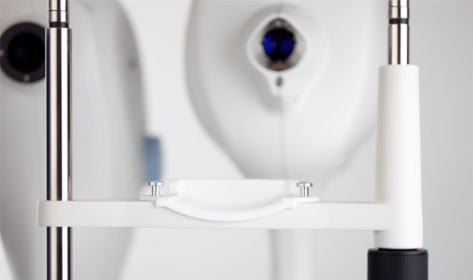
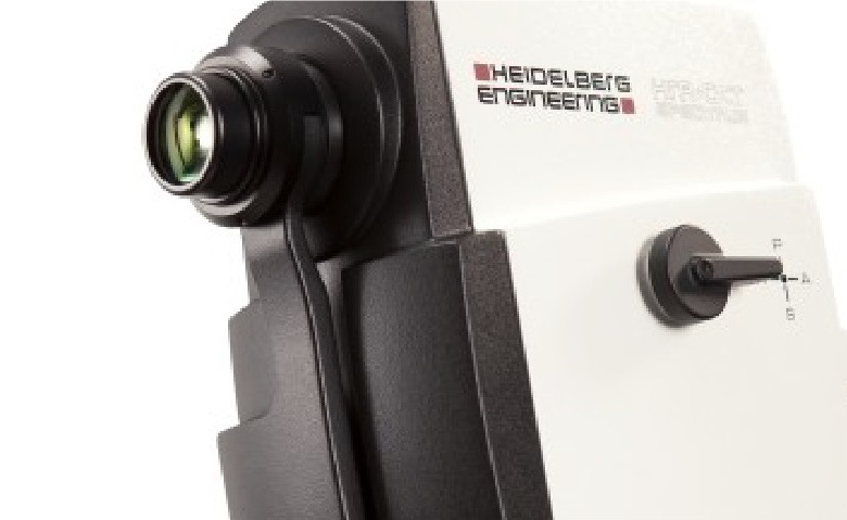
Spectralis-OCT
Spectralis-OCT technology is an optical coherence tomography imaging system that provides specialists with unique views of the structure and function of the eye. It is a non-invasive and contactless multimodal platform that provides extensive detail and information about the patient in just a few minutes. A multifunctional piece of equipment for studying disorders related to both the retina and the optic nerve, with the aim of helping ophthalmologists to obtain an early diagnosis.
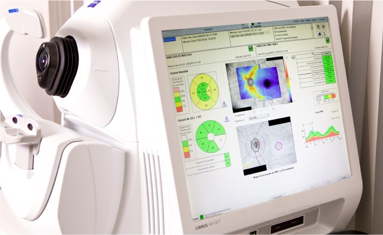
High-resolution optical coherence tomography
Optical coherence tomography (OCT) is a non-invasive that does not require pupil dilation, and it is performed by selectively scanning the retina and the optic nerve. It obtains highly detailed images of the retina and the macula in order to detect any potential anatomical changes.
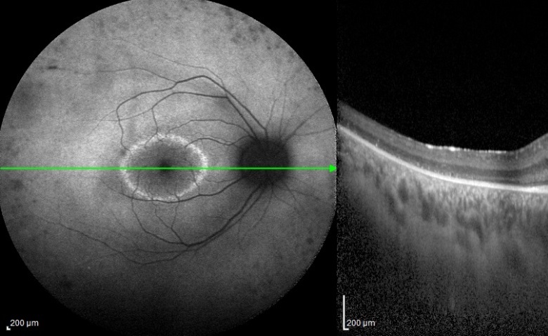
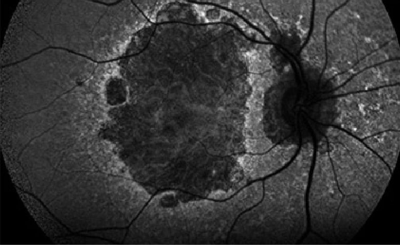
Autofluorescence imaging
Autofluorescence imaging of the fundus of the eye is a non-invasive technique that provides detailed information about the occurrence and development of complex and hereditary retinal disorders, such as AMD. It provides information about the pathophysiology of the disorder and helps ophthalmologists to better understand the nature of macular and retinal disorders so they can make more accurate diagnoses.
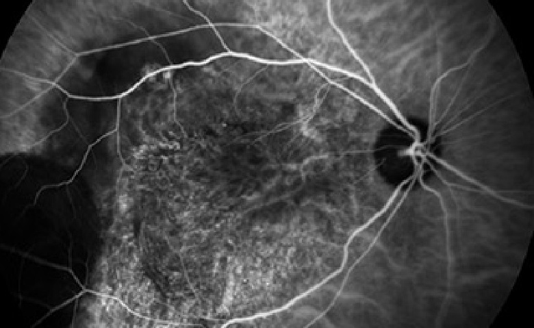
Angiography with contrast material
Angiography with contrast material is a diagnostic procedure for studying retinal vascularisation.
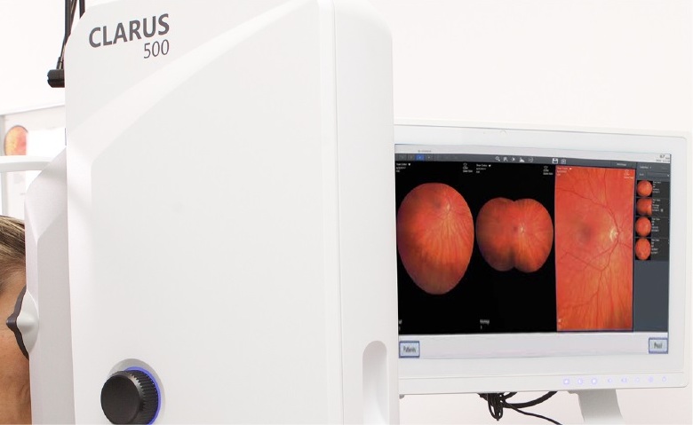
Retinograph (Clarus 500)
With this type of technology, retina specialists are able to take photographs of the fundus with more detail and precision than with a conventional retinograph without the need to dilate the pupil.
Advice for looking after your retinas
→ Annual ophthalmology check-ups.
You should see a specialist retinal ophthalmologist at least once a year. This specialist can carry out diagnostic examinations using advanced imaging technology (OCT) to prevent, manage and monitor your illness, should you be suffering from some disorder related to the retina, macula or vitreous humour. Consult with our retinal and macular specialists at Oftalvist.
→ Make sure your environment is properly lit.
→ Protect your eyes from the harmful effects of the sun.
→ Avoid rubbing your eyes.
→ Follow a healthy diet.